Introduction
The evaluation of a patient with a cavus or cavovarus foot begins with reviewing the patient’s history. Frequently, patients are unaware of a cavus foot and may present for a manifestation of a hereditary sensory motor neuropathy (e.g., Charcot-Marie-Tooth disease). In the United States, hereditary sensory motor neuropathy is the most common cause of cavovarus foot deformity. Often, an identifiable cause or family history is unknown and the diagnosis is idiopathic cavus foot.
The examination commences with an overall evaluation of the following:
- Family history of high arches
- Any systemic illness
- Any feeling of muscle weakness or tripping
- A sense of ankle instability or giving way of the ankle
- Activities of daily living, particularly occupation and athletics
- Location of discomfort, pain, or symptoms located in the foot
- Types of treatment attempted (e.g., surgery, orthosis, or braces)
The examination focuses on ambulation, standing, and an appreciation of the shape of the foot. Particularly, the examiner must determine any muscle weakness. The cavus foot represents a primary imbalance between the peroneus brevis muscle and the posterior tibial muscle, and the peroneus longus muscle and the anterior tibial muscle.
The next part of the examination focuses on determining muscle strength. The examiner must be precise and document each muscle and muscle group. Keep in mind the muscles that are phasic and those that are nonphasic. Phasic muscles include all of the flexors including the posterior tibial, flexor hallucis, flexor digitorum, and peroneals. Nonphasic muscles in the anterior compartment include the anterior tibial, extensor hallucis, and extensor digitorum longus. Knowing the location of these muscles is important when planning tendon transfers.
Next, the evaluation focuses on the severity of the cavus deformity. Is the plantar fascia tight? Where in the forefoot is the deformity? Is the first metatarsal severely plantarflexed? Is the pressure only under the first metatarsal, or is this a more global forefoot cavus that involves all five metatarsals?
The examiner must also determine if the heal is in varus. A Coleman block test is used to determine if the subtalar joint is flexible. Either a 0.5-in or 1-in block is placed under the first metatarsal, and the examiner notes the flexibility of the hindfoot and if the calcaneus is able to evert as the first metatarsal is dorsiflexed. This has an implication regarding treatment.
Are the patient’s toes clawed? If the anterior tibial muscle is weak, the common extensors begin to overact and will attempt to dorsiflex the ankle. This action, combined with the fact that often the instrinsics are weak in the cavus foot, leads to the typical claw hallux and lesser toe deformity (Slide 1). Is the ankle unstable? The cavus foot has a varus thrust on the hindfoot and ankle that leads to ankle instability.
Radiographic Examination
The radiographs must be obtained with bilateral weight bearing views of both feet and ankles. I routinely obtain stress views of the ankle, particularly the inversion stress view, to determine whether ankle instability is present. A long axial calcaneal view is also obtained to determine the extent of varus deformity of the calcaneus. On the lateral radiograph, note the apex of the cavus deformity, the calcaneal pitch angle, the talar calcaneal angle, and the talus first metatarsal angle.
Surgical Reconstruction
The reconstruction of the cavus foot depends on two factors. First, the extent of the instability on the hindfoot associated with muscle weakness, and second, the extent or rigidity of the deformity. As a general rule, surgeons should avoid arthrodesis procedures. There may be temptation to perform a triple arthrodesis with a cavus foot, but this procedure is invariably unsuccessful. A triple arthrodesis does not correct the deformity and may result in marked stress on the ankle and ultimate degenerative arthritis. Muscle balancing must be performed as well as any hindfoot osteotomy or arthrodesis.
The standard approach to correct a cavus deformity is a calcaneal osteotomy in conjunction with a plantar fascia release (Slide 2). Surgeons may also need to perform an osteotomy of the medial forefoot, either a closing wedge osteotomy of the medial cuneiform or the first metatarsal depending on the apex of the deformity.
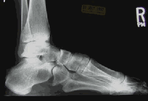
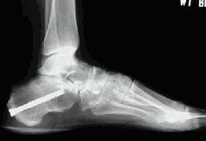
The calcaneus cannot be corrected until the plantar fascia is released. The plantar fascia release must be done immediately. The release of plantar fascia is often not sufficient because the abductor hallucis fascia is also contracted and must be released. A complete plantar fascia release is performed posteriorly. Once this is done, the calcaneal osteotomy is performed.
A triplane posterior calcaneal osteotomy is performed. This combines the elements of a standard wedge osteotomy (Dwyer) with a Samilson osteotomy that dorsally translates the calcaneus (Slide 3). The triplane osteotomy changes the pitch angle by shifting the calcaneus in a cephalad direction, removes the varus deformity with a laterally based wedge, and shifts the calcaneus laterally. Once this procedure is done, the severity of the deformity is evaluated.
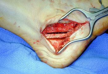
For mild deformities, the calcaneus osteotomy may be sufficient to obtain correction. For moderate deformities with the apex at the metatarsocuneiform joint, a dorsiflexion osteotomy of the medial cuneiform or first metatarsal will correct the plantarflexed deformity of the first ray (Slide 4). For a deformity that is more severe, the apex of the deformity is more proximal in the region of the navicular cuneiform joint, and an osteotomy through the entire tarsal joint (Jappas) is performed. The osteotomy is performed in an oblique manner through the cuboid to dorsiflex the cuboid and elevate the lateral column at the same time removing a wedge from the apex of the deformity.
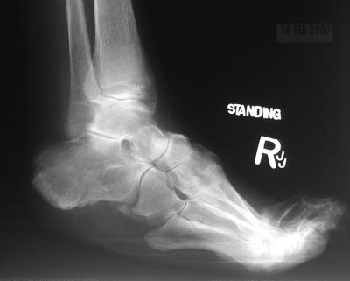
After the osteotomy is performed, one should focus on muscle balancing. Surgeons must never rely on the osteotomy alone or even an arthrodesis in the presence of muscle imbalance. The tendon transfers are performed according to the pattern of muscle weakness. A commonly used tendon transfer is a peroneus longus to the peroneus brevis tendon. This transfer removes the deforming force of the peroneus longus and increases the valgus around the hindfoot. In the presence of a foot drop, a posterior tibia tendon transfer through the interosseous membrane to the dorsal aspect of the foot is a reliable procedure. The extensor tendons are inserted into either the middle or lateral cuneiform or the cuboid depending on the severity of the deformity and extent of muscle weakness.
Additional tendon transfers may be performed using the common extensors and the extensor hallucis longus. If clawing of the forefoot is present, then the extensor hallucis longus is transferred to the first metatarsal and the common extensors are transferred to the middle cuneiform. At the same time, an arthrodesis of the hallux and lesser toe interphalangeal joints are performed. For inflexible toe deformities, a flexor to extensor tendon transfer is recommended. This increases the plantarflexion moments across the metatarsal phalangeal joint and counteracts the intrinsic weakness that perpetuates the deformity.
The ankle is evaluated and if instability is noted either clinically or radiographically, then an ankle ligament reconstruction is performed using the peroneus brevis. The Myerson modification of the Chrisman-Snook procedure is useful. If the peroneus brevis is not functional, then either half or the entire tendon may be used as part of this reconstruction.