Pathophysiology
External blows that cause tissue damage and vascular disruption lead to muscle contusions. An external blow creates a compression wave that travels through soft tissues and causes damage to the deep portion of muscle (vastus intermedius) as the external blow crushes the muscle against the adjacent bone.1,2 Extravasation of blood occurs within 24 hours, and bleeding is dispersed within and between muscle.3,4 By 1 week, acute inflammation results in elimination of debris and granulation tissue replaces the hematoma and necrotic muscle.3,4
Myoblastic activity increases and, by 3 weeks, dystrophic calcium and intramembranous bone can form in muscle and eventually ossify (Slide 1).3,4 A peripheral zone of lamellar bone, an osteoid middle zone, and a central proliferation of granulation and fibrous tissue is characteristic of myositis ossificans.2
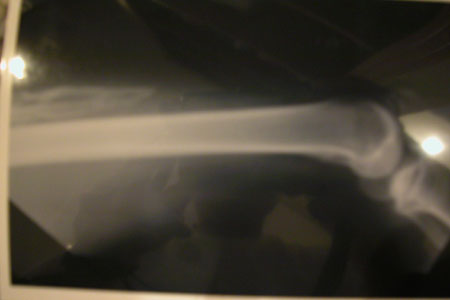
Forceful manipulation and contusion can individually lead to the development of ossification as shown in animal models.4 Despite these findings, early mobilization is beneficial after a contusion. A study comparing mobilized to immobilized muscles after muscle contusion showed that mobilized muscle returned to normal more rapidly, and histology revealed more extensive muscle regeneration in mobilized muscles. There is a beneficial middle ground with regard to mobilization of muscles after contusion.
Classification
Quadriceps contusions are graded as mild, moderate, or severe. A mild quadriceps contusion is when a patient has more than 90° of the normal knee range of motion. Forty-five degrees to 90° of normal knee range of motion is considered a moderate contusion, and less than 45° of normal knee range of motion is considered a severe contusion.2 A quadriceps contusion must be classified at 12 to 24 hours following injury. Patients with severe contusions are likely to develop myositis ossificans. 3,5,6
Myositis ossificans is classified into four types based on radiographic appearance.5-7 There can be a connection to the adjacent bone by a stalk, a periosteal type with total continuity between the heterotopic bone and native bone, a broad-based type with a portion of ectopic bone projecting into the quadriceps, and heterotopic bone without any connection to the adjacent bone.5-7 Like many classification systems, this classification is descriptive only and does not carry any prognostic significance.
History and Physical Examination
The typical history usually involves an athlete who is struck on the anterior or lateral thigh. The patient will experience pain with decreasing ability to bear weight and use the affected extremity. Occasionally, an athlete with a mild contusion will continue some level of sports participation, but there is often increasing disability within minutes to hours after injury. Patients evaluated shortly after the injury may have decreased knee range of motion, swelling, warmth, and ecchymosis. Patients with severe contusions can present with a sympathetic knee effusion or an extensor lag.
Sometimes a patient will not present for several days and a palpable mass may be noted. If radiographs are taken at this time, then they will be negative. After 3 to 6 weeks, radiographs will reveal any heterotopic ossification deep within the vastus intermedius adjacent to bone.3,5-7 Patients will typically recover with decreasing pain and maturation of the ossification followed by some degree of resorption of the myositis ossificans.3,5,6
A differential diagnosis for patients who present with a mass and radiographs revealing heterotopic ossification in the thigh includes benign lesions such as osteochondroma, osteomyelitis, and fibrodysplasia ossificans progressiva. Malignant lesions, such as osteosarcoma, chondrosarcoma, and synovial cell sarcoma, must also be considered. Most of these diagnoses can be ruled out based on the radiographic appearance of the lesion, family history, physical examination, and associated constitutional symptoms.
A history of trauma does not rule out a malignant process as 40% of patients with osteosarcomas will present with a history of trauma to the affected area.2,7
Management of Quadriceps Contusion and Early Myositis Ossificans
Rest, cooling, compression, and elevation are beneficial. Early aggressive range of motion and recurrent injury are reported to increase disability and increase incidence of myositis ossificans.3-6 Although forceful manipulation may be detrimental, some form of early mobilization is beneficial.
Studies have shown that the rates of myositis ossificans increase with injury severity.3,5,6 The incidence of myositits ossificans ranges from 0% to 9% for mild quadriceps contusions and 17% to 72% for moderate to severe quadriceps contusions.3,5,6
Using a specific postinjury protocol, two studies reported a 17% to 20% incidence of myositis ossificans.3,5 This particular protocol consisted of limiting hemorrhage, restoration of motion, and functional rehabilitation focusing on achieving early extension. A follow-up study employed a modified postinjury protocol that focused on early flexion instead of extension.6 This modification resulted in a 9% rate of myositis ossificans and decreased disability time by 66% for moderate contusions and 71% for severe contusions. No aspirations or nonsteroidal anti-inflammatory medications (NSAIDs) were used in this study. No patient who developed myositis ossificans had late symptoms, and all patients returned to preinjury levels of sporting activities.
The role of ultrasound after quadriceps contusion is not well documented. It is advisable that any modality that produces heat should be avoided in the early treatment of muscle contusions.
The role of NSAIDs, low-dose irradiation, or steroids is not extensively described in the literature. Irradiation and NSAIDs have been shown to prevent formation or recurrence of heterotopic ossification after total joint replacement and excision of heterotopic ossification.8,9 NSAIDs, including Naproxen (750 mg qd) or indomethacin (50 mg bid), are recommended for 2 to 6 weeks.8,9 Although corticosteroids and single-dose anabolic steroids have been reported to be effective for healing muscle contusions in a rat model, histology has revealed disorganized muscle fiber regeneration.10 The role for steroids in human muscle contusions is unclear and requires further research.
The development of myositis ossificans does not correlate with late functional disability in the majority of patients.2,3,5-7 If the ossification persists and is associated with muscle atrophy, joint limitation, and pain, excision is recommended only after maturation of the ossification.2,5-7 Despite excision, recurrence rates of up to 67% have been reported.7
Three-Phase Management for Quadriceps Contusions
Phase one involves rest, compression, cooling, and elevation for 24 to 48 hours regardless of the injury severity. Cooling and compression can be done with ice bags and compression bandages or cryocuff wraps. Patients with moderate to severe quadriceps contusions may also benefit from 2 to 6 weeks of NSAID treatment. Occasionally, patients with severe quadriceps contusions require hospitalization for pain management. I recommend positioning the hip and knee in a degree of flexion that does not increase pain severity (Slide 2).
Phase two consists of restoring mobility to the affected extremity when patients are pain free at rest. This consists of gentle active and passive range of motion exercises focusing on regaining flexion. When just beginning to mobilize patients and for several months postinjury, padding the affected area may be helpful in avoiding a reinjury. Weight bearing, as tolerated, is allowed and continued compression is maintained. Walking, cycling, and jogging are allowed if no additional pain is experienced with these exercises. If increasing pain or reinjury occurs, then patients are sent back to the previous phase.
Phase three consists of functional rehabilitation. After regaining nearly full range of motion and near normal strength of the knee, patients return to sport-specific activities. Rehabilitation is not altered if myositis ossificans develops, but a confirmational radiograph is taken if a palpable mass is noted. If the mass is associated with muscle atrophy, limited joint motion, and pain after conservative treatment, excision after maturity of the lesion is recommended.
References
- Ackerman LV. Extra-osseous localized non-neoplastic bone and cartilage formation (so-called myositis ossificans): Clinical and pathological confusion with malignant neoplasms. J Bone Joint Surg Am. 1958; 40:279-298.
- Fu FH, Stone DA, eds. Sports Injuries: Mechanisms, Prevention, Treatment. Baltimore, Md: Williams & Wilkins; 1994.
- Rothwell AG. Quadriceps hematoma: A prospective clinical study. Clin Orthop. 1982; 171:97-103.
- Michelsson JE, Granroth G, Andersson LC. Myositis ossificans following forcible manipulation of the leg: A rabbit model for the study of heterotopic bone formation. J Bone Joint Surg Am. 1980; 62:811-815.
- Jackson DW, Feagin JA. Quadriceps contusion in young athletes. J Bone Joint Surg Am. 1973; 55:95-105.
- Ryan JB, Wheeler JH, Hopkinson WJ, et al. Quadriceps contusions: West Point update. Am J Sports Med. 1991; 19:299-304.
- Delee JC, Drez D, Stanitski CL, eds. Orthopaedic Sports Medicine: Principles and Practice. Philadelphia, Pa: WB Saunders; 1994.
- Vielpeau C, Joubert JM, Hulet C. Naproxen in the prevention of heterotopic ossification after total hip replacement. Clin Orthop. 1999; 369:279-288.
- Kienapfel H, Koller M, Wust A, et al. Prevention of heterotopic bone formation after total hip arthroplasty: A prospective randomized study comparing postoperative radiation therapy with indomethacin medication. Arch Orthop Trauma Surg. 1999; 119:296-302.
- Beiner JM, Jokl P, Cholewicki J, et al. The effect of anabolic steroids and corticosteroids on healing of muscle contusion injury. Am J Sports Med. 1999; 27:2-9.