Introduction
The Achilles tendon is the strongest tendon in the body. During ambulation the Achilles tendon is subjected to high tensile loads that commonly result in its injury. Pathological changes to the Achilles tendon occur due to two distinct conditions. First is the noninsertional Achilles tendonitis, most commonly an overuse phenomenon that occurs 2 cm to 6 cm proximal to the calcaneal insertion and constitutes the most frequent and debilitating condition in active runners. It was reported in 6.5% to 18% of active runners, mostly men. Among older male athletes, these injuries occur at a higher rate than do most other overuse injuries.1
By contrast, posterior heel disorders (e.g., insertional Achilles tendonitis, retrocalcaneal bursitis, Haglund deformity and pretendon bursitis) are much less common conditions estimated to occur in 9% of patients presenting with Achilles tendon overuse injuries.2 This constellation of symptoms usually presents in combination. The patient population is usually older, poorly conditioned athletes or workers in occupations requiring prolonged standing or walking. There is also a subgroup of overweight, sedentary individuals.3
Anatomy
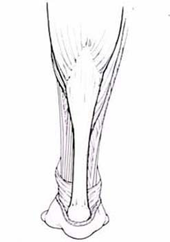
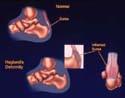
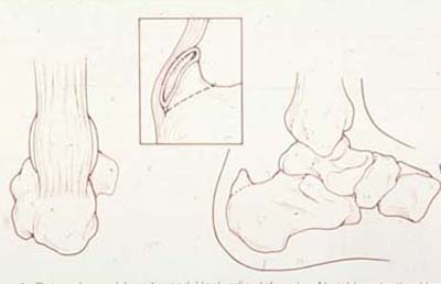
Three muscle bellies, the medial and lateral gastrocnemius heads arising from the distal femoral condyles and the soleus muscle from the posterior aspect of the tibia and fibula, converge to form the "triceps surae" or the Achilles tendon (Slide 1). The plantaris muscle, which originates from the lateral femoral condyle, passes between the gastrocnemius and soleus bellies to reside medial to or anterior to the Achilles tendon and inserts into it or the calcaneous. Although individual ruptures may occur, it is not usually involved in tendo Achillis ruptures or other pathology. The Achilles tendon courses distally, rotates 90° internally, with the soleus contribution being medial to that of the gastrocnemius, and inserts into the middle third of the flat surface of the posterior calcaneal tuberosity. On average, the tendon starts 1 cm inferior to the top of the posterior calcaneus and covers an area that is 2 cm wide, with the medial insertion being wider than the lateral.4 A section of the tendon, 2 cm to 6 cm proximal to its insertion site, is a hypovascular zone as demonstrated by Lagergren and Lindholm.5 This zone is the narrowest in cross section and corresponds to the most common site of tendon pathology, including paratenonitis, tendinosis and tendon rupture. The tendon is surrounded by a paratenon that has a single layer of cells with variable structure, not a true tenosynovium. The retrocalcaneal bursa is a constant horseshoe-shaped bursa found anterior to the tendon insertion site, which separates it from the posterior/superior calcaneal process. The pretendon or subcutaneous calcaneal bursa lies posterior to the tendon insertion and is variably present. Haglund deformity is a prominent posterior/superior calcaneal process described by Haglund in 1928 (Slide 2).
Noninsertional Achilles Tendinitis
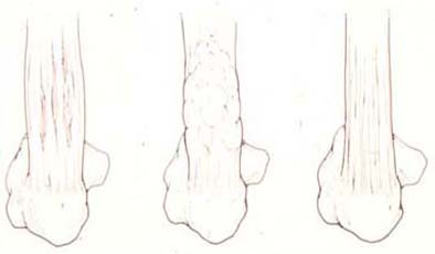
Considered by some authors to be the most common ailment seen in sports medicine clinics, Achilles tendinitis constitutes 10% of all lower extremity complaints in runners.6, 7 Several factors contribute to development of these overuse injuries including age, degeneration of the tendon, areas of decreased vascularity, malalignment, especially genu varus and cavus foot deformity.8 Also, changes in training habits, such as sudden increase in mileage or change in training intensity or training surfaces, can contribute to overuse injuries.6 Puddu et al9 presented an anatomic classification of noninsertional Achilles tendonitis (Slide 3) into an inflammation of the lining of the tendon or paratenonitis, or paratenonitis with tendinosis, i.e., intratendinous degeneration, and finally tendinosis or noninflammatory intratendinous degeneration due to aging, microtrauma or vascular compromise.
Clinical Presentation
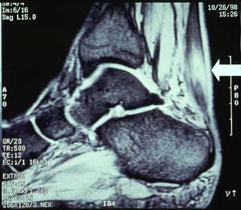
A history of gradual progressive pain and swelling, 2 cm to 6 cm proximal to the tendon insertion, is typical for an older male athlete presenting with normal plain calcaneus radiographs.10 The pain duration and intensity progresses, at first only experienced with prolonged activity, stair climbing, and later progressing to pain with daily living activities. Careful examination may help define the stage of the disease. In acute paratenonitis alone there is localized swelling and tenderness, which then become more diffused. The tender area is usually immobile. In paratenonitis with tendinosis, there is an area of thickening and irregularity to palpation that presents along with increased tenderness deep within the tendon. Most importantly, the area of tenderness and swelling moves as the ankle is dorsiflexed and plantarflexed. Finally, in isolated tendinosis the patient presents with no pain or minimal intermittent pain. A palpable nodule or irregularity may be felt at the area of degeneration that moves with excursion of the tendon. This nodule is usually asymptomatic. Partial and complete rupture may follow, often with a trivial injury. Ultrasonography and, more recently, magnetic resonance imaging examinations (Slide 4) are utilized in preoperative planning to delineate the extent of tendinosis and/or rupture in the tendon substance.11
Treatment
Nonoperative treatment is recommended in most cases presenting with paratenonitis with or without tendinosis. Nonsteroidal anti-inflammatory drugs (NSAIDs), Achilles tendon stretching, and a 5/16 of an inch heel lift are prescribed (Slide 5). We now use an elastic heel gauntlet which has a built in heel lift padding on either side of the tendo Achillis, which provides compression, protection and less stress on the tendon.
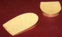
The mainstay of treatment is activity modification as follows:
Patients with acute inflammation and significant pain may require 7 to 10 days of immobilization in a short-leg cast or a walking boot with the heel elevated, followed by an active therapy program focused on strengthening of the calf muscles and stretching of the Achilles tendon. A gradual return to running activities is begun with emphasis on increasing distance and intensity without the return of pain. Orthotic devices are prescribed, especially in runners with excessive pronation.1 With these modalities, 90% to 95% of patients will resolve their symptoms.12 The authors occasionally use iontophoresis or phonophoresis for transcutaneous delivery of anti-inflammatory medications but strongly advise against the use of direct steroid injection based on the impression that it may enhance tendon attrition and rupture.
Operative treatment is reserved for recalcitrant cases resistant to an adequate, nonoperative treatment for 12 weeks or more.3 MRI examination is recommended in the preoperative period to better evaluate the site and extent of pathology as well as possible partial rupture. Several surgical approaches have been described in the literature.3, 12-14 In the authors’ experience, a longitudinal medial incision that avoids the lateral perforating vessels and possible injury to the posterior branch of the sural nerve is recommended. The surgical treatment depends on the condition of the tendon; in paratenonitis alone, we recommend the release and stripping of the thickened paratenon from the tendon substance and release of adhesions. If tendinosis is encountered as well, the tendon is debrided by making longitudinal incisions into its substance, excising the necrotic tissue, and the tendon is repaired with nonabsorbable suture.14 In the postoperative period, patients who had paratenonitis debridement alone can progress to early mobilization. In paratenonitis with tendinosis, protection in a weightbearing, short-leg cast or a walking boot in neutral position for 4 to 6 weeks is followed by gradual return to running activities using a heel lift in all cases. Full recovery of calf strength is slow, especially eccentric plantar flexion strength, which may take up to 1 year.11
Results
Reviewing the literature, the results of operative treatment produced good-to-excellent results in 86% of patients with paratenonitis alone, while paratenonitis and tendinosis requiring tendon debridement achieved 73% good to excellent results.15
Posterior Heel Disorders
Considered a separate entity from noninsertional Achilles tendinitis, posterior heel disorders are 4 distinct conditions: insertional Achilles tendinitis, retrocalcaneal bursitis, Haglund deformity and pretendon bursitis. These conditions affect a more diverse segment of the population including younger active runners, usually presenting with localized disease, as well as an older, less active and often overweight patient with diffuse involvement and often a poorer prognosis.3 Haglund triad (Slide 6) of insertional tendinitis, retrocalcaneal bursitis and Haglund deformity occurring in combination seems to be the most common presentation accounting for 20% to 30% of all Achilles tendinitis cases.3,16
Pathophysiology
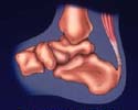
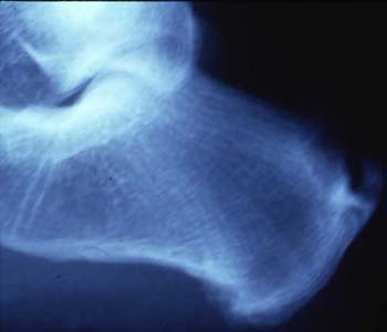
Insertional Achilles tendinitis is often the earliest condition to manifest (Slide 7). It is an attritional, degenerative condition caused by chronic overuse, i.e., an enthesopathy. On histopathological examination, changes similar to the more proximal tendinosis are seen. The paratenon, however, does not become as thickened or as obviously involved. With continued overuse, abnormal traction on the posterior calcaneal tuberosity results, which produces reactive changes and later ossification within the insertion of the tendon or anterior and lateral to it (Slide 8). This bony projection, frequently referred to as "fishhook" osteophyte, can be quite painful with continuing pressure and shoe wear irritation. Retrocalcaneal bursitis and Haglund deformity often coexist with the above clinical picture. Often seen predisposing biomechanical abnormalities include a rigid plantar flexed first ray, hindfoot varus, and, on occasion a tight Achilles tendon. These abnormalities can lead to increased shear stresses at the osseous soft tissue interface along the posterosuperior aspect of the calcaneus.17 The result is an irritation to the bursa, more fluid secretion and increased impingement on the anterior Achilles tendon, especially with ankle dorsiflexion. Haglund deformity, a prominence of the posterosuperior calcaneal tuberosity, further compresses the bursa against the insertion of the tendo Achillis with dorsiflexion and exacerbates the "nut-cracker" effect on the bursa. A tight heel counter or uphill running with continuous forced ankle dorsiflexion poses an added challenge to the posterior heel impingement.
Differential Diagnosis
When evaluating posterior heel pain, one should consider other entities in the differential diagnosis, especially systemic inflammatory diseases. Seronegative spondyloarthropathies, gout, familial hyperlipidemia, sarcoidosis, diffuse idiopathic skeletal hyperostosis (DISH), and reaction to drugs such as systemic corticosteroids or oral fluroroquinolones, have also been cited in cases of Achilles tendinitis or retrocalcaneal bursitis.18-22 The overall incidence of retrocalcaneal bursitis with rheumatoid arthritis is estimated to be between 2% and 10%.23
Diagnostic Studies
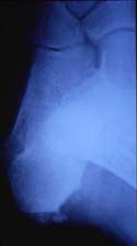
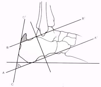
Plain radiographs of the calcaneus will show evidence of heterotopic ossification with bone attached to the posterior calcaneus (fishhook osteophytes) and extending proximally within the tendon just above its insertion (Slide 8). An oblique foot projection is necessary to display the intratendinous ossification in multiple areas that may be obscured on a direct lateral view (Slide 9). Haglund deformity is a characteristic prominent posterosuperior calcaneal tuberosity evident on a direct lateral view of the calcaneus and best measured by the calcaneal parallel pitch lines (Slide 10). This reproducible measurement is a line drawn starting from the posterior lip of the talar articular facet and parallel to a tangent line drawn along the plantar aspect of the calcaneus. Any posterosuperior calcaneal tuberosity bone that is superior to this line is excessive and represents a Haglund deformity. Although an MRI evaluation is not necessary to diagnose or treat posterior heel disorders, it is beneficial in the preoperative evaluation to determine the extent of tendon degeneration, the debridement required and the possible need for a more extensive tendon reconstruction.
Treatment
The nonoperative treatment of the posterior heel painful triad is a trial of NSAIDs, a 5/16 inch heel lift, and Achilles tendon stretching. Exercise modification and Achilles tendon strengthening follows as the pain subsides. Iontophoresis or phonophoresis is often helpful. On rare occasions immobilization in a walking boot with a heel lift or an injection of steroids into the retrocalcaneal bursa is helpful. We also use the elastic gauntlet with the built in heel lift for this entity.
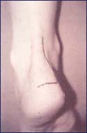
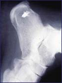
With failure of adequate nonoperative treatment, operative intervention is indicated. With the patient prone, the authors approach the area of pathology through a medial longitudinal and inferior transverse J incision (Slide 11). In the authors’ opinion, such an incision preserves the lateral venous outflow and avoids the hypersensitivity produced by scarring or injury to the posterior branch of the sural nerve. The inferior limb is just distal to the tendon insertion; it is important to maintain a full-thickness subcutaneous flap to avoid wound slough. Longitudinal incisions are made in the tendon to remove degenerated tendon, intratendinous heterotopic bone, and any calcaneal bony spurs. If the insertion is disrupted mid-portion, or a third or more of the tendon is detached during debridement, a tendon reattachment is performed with bone suture anchors (Slide 12 and Slide 13). If the entire tendon is involved and detachment is necessary, reinforcement with local tendon graft (plantaris), or a tendon transfer into the calcaneal tuberosity — most commonly of the flexor hallucis longus (FHL), is indicated. Den Hartog6 has suggested frequent use of the FHL to reinforce a frayed tendo-Achillis, using a simple harvest technique from the same wound. The retrocalcaneal bursitis and Haglund deformity is approached from the medial aspect; the bursa is excised along with the adjacent anterior paratenon. The Haglund deformity is excised in an oblique direction down to the area directly above the actual tendon insertion (Slide 12). Intraoperative fluoroscopy or plain radiographs are necessary to determine the adequacy of bone excision.
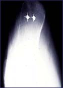
Postoperative management requires the application of a short-leg cast in slight equinus. In cases of tendon reattachment or tendon transfer the patient is protected in cast for 6 weeks, nonweight-bearing, followed by gradual return to ambulation in a walking boot with a heel lift for 2 more weeks, followed by use of the lift for another month (3 months total). For debridement alone immobilization in a walking boot with a heel lift is used for up to 4 weeks, followed by heel lift alone in a shoe for 2 weeks (6 weeks total).
Postoperative Results
The overall success rate mentioned in the literature is 85% to 97% good to excellent results.3, 24, 25 The postoperative rehabilitation program time needed prior to return to activity depends on the integrity of the remaining Achilles tendon and its insertion. Complications, such as wound slough and infection with postoperative rupture are serious problems that require aggressive debridement, often complex tendon reconstruction, and bone tissue vascularized soft tissue coverage. Obviously, skin flap care is essential, and prompt treatment of even a minor slough may avoid the more disastrous complication. Rupture alone is a simpler matter. Reinsertion, reinsertion with a proximal Z lengthening, and/or reinforcement with a tendon transfer are all considerations.
Pretendon Bursitis
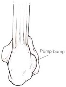
A pretendinous bursitis may occur between the posterior heel skin and the underlying tendoAchillis, especially a prominent one which contains heterotopic bone. This "fourth entity" of the Haglund triad is treated with the other components and at surgery is excised first. An additional type of bursitis may arise just lateral to the tendo-Achillis insertion, and is known as a "pump bump." This common condition is largely associated with female pump-style shoes. A stiff heel counter compresses the pretendon bursa against the prominent calcaneal posterolateral prominence with resultant inflammation and enlargement (Slide 14). Over time, more bone may be formed under the bursa, owing to the irritation.
Clinical Presentation
Patients complain of pain at the enlarged posterolateral calcaneal prominence. They note difficulty wearing fashionable shoes and that the bump is slowly enlarging. On physical examination, focal swelling and tenderness along with a palpable bony prominence is noted just lateral to the Achilles tendon. The Achilles tendon itself and the retrocalcaneal bursa are not involved. Radiographs, especially calcaneal axial views, will show the bony prominence, but otherwise are unremarkable.
Treatment
Patients are encouraged to use shoes with a softer heel counter in an effort to relieve the external compression on soft tissue and bone. The silopad Achilles heel pad (silipos) is effective in protecting the inflamed tissue. A trial of NSAIDs is indicated. On occasion, operative intervention is indicated for a patient with chronic persistent symptoms. A lateral longitudinal incision, with care taken to protect the sural nerve, is made. The bursa and any posterolateral bone prominence are excised. Postoperatively, weightbearing and shoe wear is permitted once the soft tissue is healed.
References
- Kvist M. Achilles tendon injuries in athletes. Sports
Med. 1994; 18:173-201.
- Schepsis AA, et al. Surgical management of Achilles tendon
overuse injuries. Am J Sports Med. 1994; 22:611-619
- DiGiovanni BF, Gould JS. Achilles tendinitis and posterior
heel disorders. Foot Ankle Clin. 1997; 2(3):411
- Chao W, et al. Achilles tendon insertion: An in vitro
anatomic study. Foot Ankle Int. 1997; 18:81-84.
- Lagergren C, Lindholm A. Vascular distribution in the
Achilles tendon: An angiographic and microangiographic study. Acta Chir
Scand. 1958/1959; 116:491-495,
- Den Hartog B. Presentation on Achilles tendinitis. AOFAS
Annual Summer Meeting. Boston, MA; 1998.
- James S, et al. Injuries to runners. Am J Sports
Med. 1978; 6:40-49.
- Jones DC, James SL. Partial calcaneal osteectomy for
retrocalcaneal bursitis. Am J Sports Med. 1984; 12:72-73.
- Puddu G, et al. A classification of Achilles tendon disease.
Am J Sports Med. 1976; 4:145-150.
- Hakan A, et al. Chronic Achilles tendinitis and calf muscle
strength. Am J Sports Med. 1996; 24(6):829.
- Hakan A, et al. Achilles tendinosis and calf muscle
strength: The effect of short-term immobilization after surgical treatment.
Am J Sports Med. 1998; 26(2):166.
- Clancy WG, Heiden EA. Achilles tendinitis, treatment in the
athlete. Foot Ankle Clin. 1997; 2(3):429-438.
- Clement DB, et al. Achilles tendinitis and paratendinitis
etiology and treatment. Am J Sports Med. 1984; 12:179-184.
- Gould N, Korson R. Stenosing, tenosynovitis of the pseudo
sheath of the tendo-Achilles. Foot Ankle. 1980; 1:179-187.
- Nelen G, et al. Surgical treatment of chronic Achilles
tendonitis. Am J Sports Med. 1989; 17:754-759.
- Haglund P. Beitrag zur uliwik der achilesse have. Z
Orthop Chir. 1928; 49:49.
- Jones DC. Achilles tendon problems in runners. AAOS
Instructional Course Lectures. 1998; 47:419-427.
- Crowder SW, Jaffrey UA. Sarcoidosis presenting as Achilles
tendinitis. J R Soc Med. 1995; 88:335-336.
- Gerester JL. Achilles tendinitis as a severe clinical
feature of DISH (letter). J Rheumatol 1995; 22:1212-1214.
- Movin T, et al. Pathology of the Achilles tendon in
association with Ciprofloxacin treatment. Foot Ankle Int. 1997;
18(5):297.
- Scioli MW. Other arthritides. In: Scinmorro GJ, ed.
AOFAS Foot and Ankle Manual. Philadelphia: Lea & Febiger.
1991; 186-194.
- West MB, Gow B. Ciprofloxacin, bilateral Achilles
tendinitis, and unilateral tendon rupture — A case report. New
Zealand Med J. 1998; 18-19.
- Turlik MA. Seronegative arthritis as a cause of heel pain.
Clin Podiatr Med Surg. 1990; 7:369-375.
- Gerken AP, McGarvey WC, Baxter DE. Insertion of Achilles
tendinitis. Foot Ankle Clin. 1996; 1:237-248.
- Sammarco GJ, Taylor A. The operative management of Haglund
deformity in the non athlete: A retrospective study. Foot Ankle Int.
1998; 19(11):724. Presented at Winter Meeting of American Orthopaedic
Foot and Ankle Society, 1997.